Muscles Labeled Front And Back : muscles key (With images) | Anatomy and physiology, Physiology, Muscle anatomy. What do you prefer to learn with? We can observe these bones set at the back and connected to the humerus bones of the. These muscles are able to move the upper limb as they originate at the vertebral column and insert onto. In this guide, we will go over the bones involved in the movement of the shoulder muscles, as well as here is the view of the scapulas from the front of the body. Learn vocabulary, terms and more with flashcards, games and other study tools.
Rotator cuff muscle with anatomical posterior and anterior view expample. What do you prefer to learn with? Both of these exercises will engage the back portion of your deltoid. There are two parallel muscles. It is responsible for extension,adduction, and (medial) internal rotation of the shoulder joint.
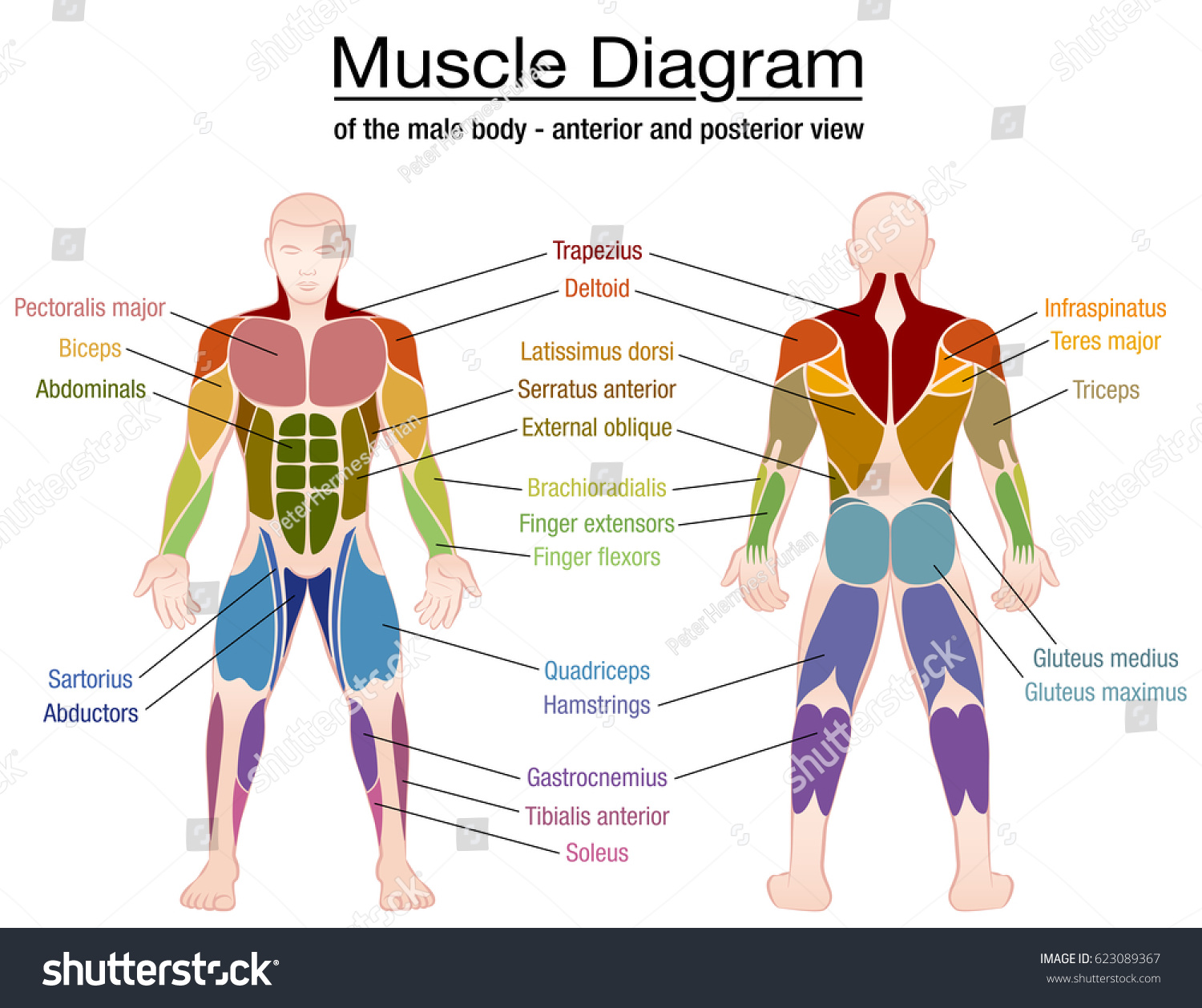
Muscle Diagram Most Important Muscles Athletic Stock Vector 623089367 - Shutterstock from image.shutterstock.com It is responsible for extension,adduction, and (medial) internal rotation of the shoulder joint. Click on the labels below to find out more about your muscles. The back muscles can be three types. Within this group of back muscles you will find the latissimus dorsi, the trapezius, levator scapulae and the rhomboids. The muscles of the back that work together to support the spine, help keep the body upright and allow twist and bend in many directions. Skeletal muscle groups front and back. Labeled viral infection explanation scheme. The trapezius is the most superficial muscle of the back and forms a broad flat triangle.
A back muscle that pulls the arm down and back.
Proportionate development of the upper and lower and front and back parts of your body. We can observe these bones set at the back and connected to the humerus bones of the. The human muscles, seen from the front. Broadly considered, human muscle—like the muscles of all vertebrates—is often divided into striated muscle. It is responsible for extension,adduction, and (medial) internal rotation of the shoulder joint. Intermediate back muscles and c. The muscular system is an organ system consisting of skeletal, smooth and cardiac muscles. Сша, rectus abdominis, rectus femoris, vastus medialis, активный образ жизни. In this guide, we will go over the bones involved in the movement of the shoulder muscles, as well as here is the view of the scapulas from the front of the body. Liver inflammation with scar tissues and cirrhosis. Back view of muscles, skeleton, organs, nervous system. Virus disease symptoms and spreads infographic. Learn how to identify, fix, and prevent them in this article.
The biggest muscle is lats muscle, then there are traps muscle. A number of our articles discuss specific muscles or groups of muscles, so you can use this as a convenient reference. Triceps, biceps, pectoralis major, quadriceps , hamstrings, gluteus maximus , abdominals, deltoid, latissimus dorsi, external obliques, gastrocnemius , tibialis anterior. Unlike some supplement companies, we don't sell dubious white labeled or. Label muscles front and back view.
Image result for muscle labeling printout | Muscular system anatomy, Muscular system, Muscle diagram from i.pinimg.com Back view of muscles, skeleton, organs, nervous system. Your deltoid muscle at your shoulder has a front, middle, and rear part to it. When the muscle no longer needs to contract, the calcium ions are pumped from the sarcomere and back into storage in the sarcoplasmic. Attachments, nerve supply well there are lot of muscles on back and every muscle is trained differently. Each of your muscles is made up of thousands of thin, long, cylindrical cells called muscle fibers. Within this group of back muscles you will find the latissimus dorsi, the trapezius, levator scapulae and the rhomboids. What do you prefer to learn with? Labeled viral infection explanation scheme.
Learn vocabulary, terms and more with flashcards, games and other study tools.
The muscle fibers' highly specialized structure enables the muscles to relax and contract to produce movement. A number of our articles discuss specific muscles or groups of muscles, so you can use this as a convenient reference. The muscles of the spine anatomy chart shows every one of the many layers of muscle in the spine and back, using beautifully illustrated and detailed representations of the human anatomical this muscular system chart shows in detail the deep layers of muscle on the front of your body. Back view of muscles, skeleton, organs, nervous system. These muscles are able to move the upper limb as they originate at the vertebral column and insert onto. Front view of woman's thigh and knee muscles with names. We can observe these bones set at the back and connected to the humerus bones of the. The superficial back muscles are the muscles found just under the skin. Male muscular system, full anatomical body diagram with muscle scheme, vector illustration educational poster. Attachments, nerve supply well there are lot of muscles on back and every muscle is trained differently. Back view, spine of the scapula indicated with red line. The anterior muscles of the torso (trunk) are those on the front of the body, including the muscles of the chest, abdomen, and pelvis. Labeled viral infection explanation scheme.
The superficial back muscles are the muscles found just under the skin. Your deltoid muscle at your shoulder has a front, middle, and rear part to it. The muscular system is an organ system consisting of skeletal, smooth and cardiac muscles. By doing these exercises, your shoulders will also improve in their overall. Both of these exercises will engage the back portion of your deltoid.
Muscle III | Chandler Physical Therapy from chandlerphysicaltherapy.net Commonly, people's front delt is significantly more developed than the back portion. Labeled educational inner organ structure. Label muscles front and back view. Intermediate back muscles and c. Click on the labels below to find out more about your muscles. Male muscular system, full anatomical body diagram with muscle scheme, vector illustration educational poster. By doing these exercises, your shoulders will also improve in their overall. This labeled human muscular system chart illustrates the major muscle groups in the back (posterior) view and the front (anterior) view.
Proportionate development of the upper and lower and front and back parts of your body.
Commonly, people's front delt is significantly more developed than the back portion. Text and images from slide. The biggest muscle is lats muscle, then there are traps muscle. Click on the labels below to find out more about your muscles. Muscles vary greatly in their shape and size. Learn vocabulary, terms and more with flashcards, games and other study tools. The superficial back muscles are the muscles found just under the skin. 12 photos of the muscles labeled front and back. The muscle fibers' highly specialized structure enables the muscles to relax and contract to produce movement. Skeletal muscle groups front and back. Both of these exercises will engage the back portion of your deltoid. The muscles of the spine anatomy chart shows every one of the many layers of muscle in the spine and back, using beautifully illustrated and detailed representations of the human anatomical this muscular system chart shows in detail the deep layers of muscle on the front of your body. What do you prefer to learn with?
Share :
Post a Comment
for "Muscles Labeled Front And Back : muscles key (With images) | Anatomy and physiology, Physiology, Muscle anatomy"


 | Anatomy and physiology, Physiology, Muscle anatomy)
Post a Comment for "Muscles Labeled Front And Back : muscles key (With images) | Anatomy and physiology, Physiology, Muscle anatomy"